Loading…Loading…

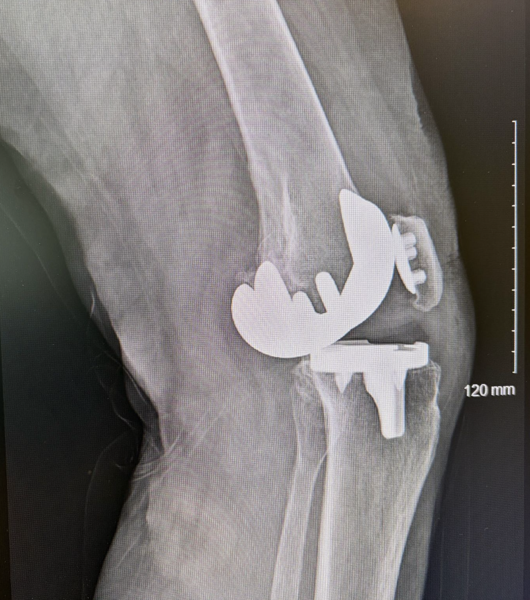
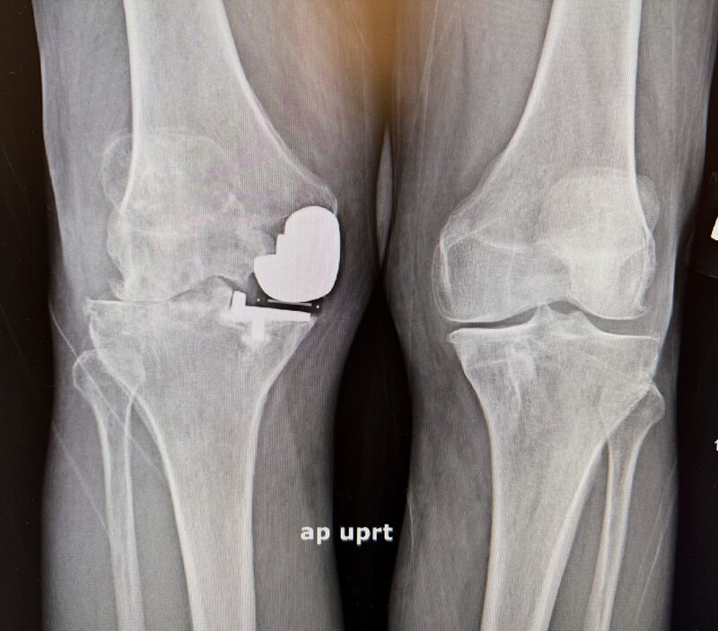
Eight years out from a TKA, and the X-ray tells the story before the patient finishes the sentence. Significant polyethylene wear. Gross instability. Infection workup negative. Cobalt and chromium both running 2x normal — the metal is involved now, not just the plastic. In my OR, this is the fork I think about hardest: with instability plus metallosis on top of wear, is an isolated poly exchange ever enough — or has the metal damage already made that decision for you? For the revision surgeons: where's your line? What pushes you from poly swap to full revision in a case like this — the instability pattern, the ion levels, what you find once you're in, or something else? More details here
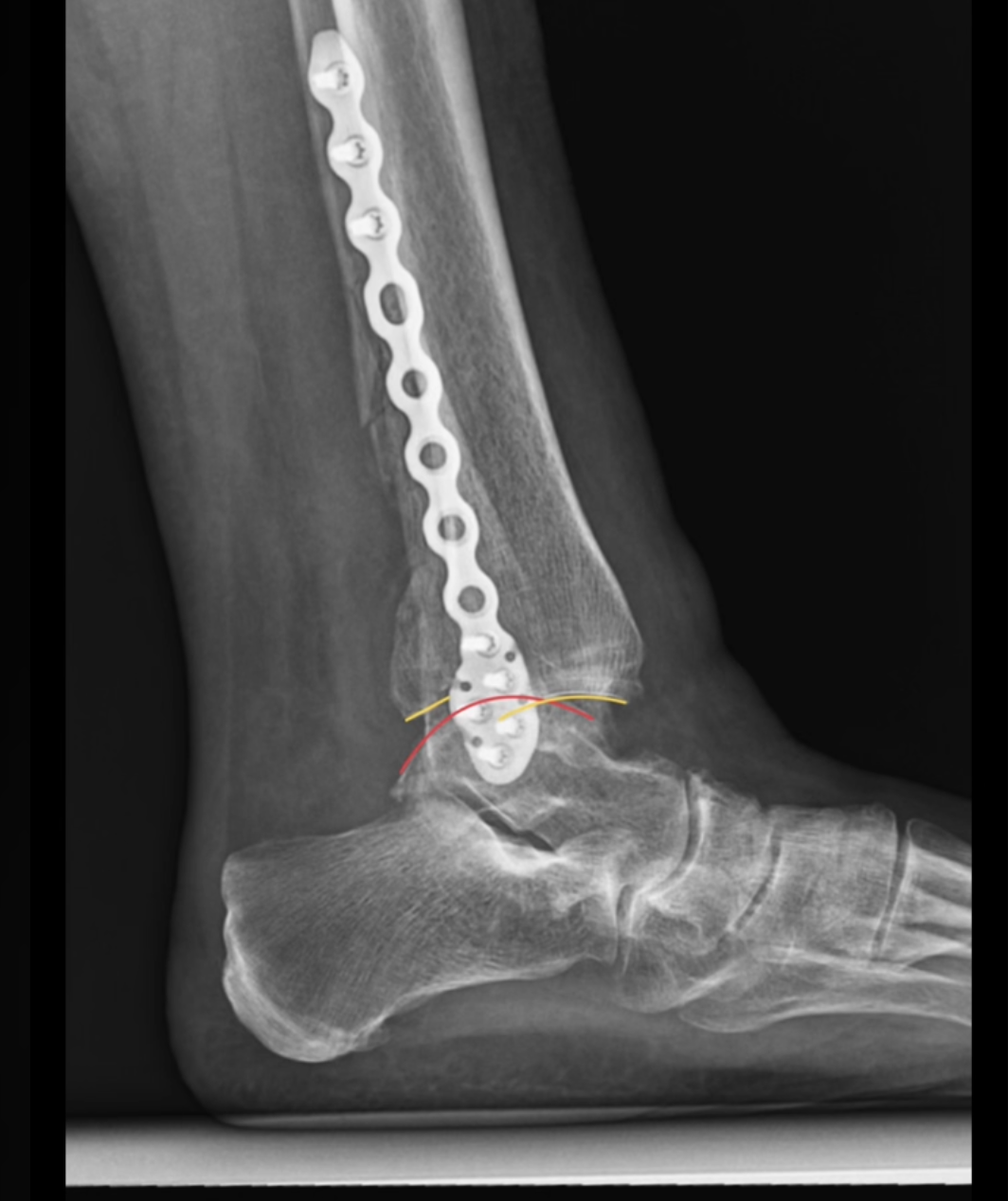
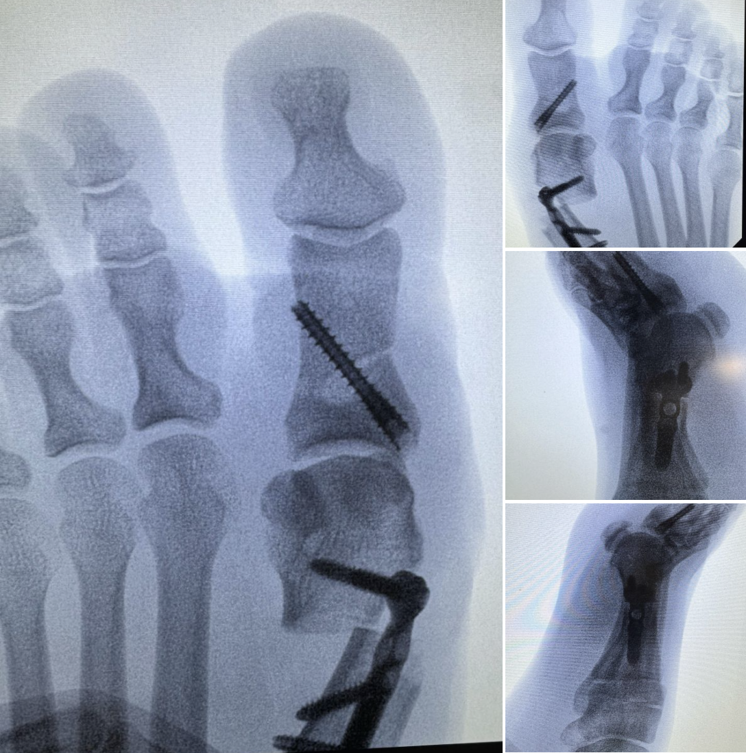
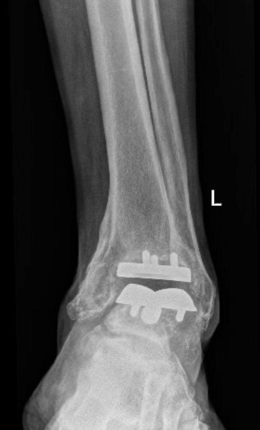
This is the case of an older patient who sustained a low-energy posterior #pilon fracture-dislocation of the ankle. The ankle was reduced in the ER and splinted. She was treated with open reduction and internal fixation, including the posterior distal tibia. The fibula was fixed with bridge fixation due to comminution. The tibia was fixed with posterior-to-anterior #TTFNs from OSSIO through an #MPM approach. After fixation, the external rotation stress was negative. At her first post-op, they were noted to be dislocated again. Upon questioning, they reported using the leg for standing and transferring in the splint, despite post-op non-weight-bearing restrictions. With these small fragments, in hindsight I could’ve used #antiglide plates. Since the joint was dislocated for an unknown amount of time, the decision was made to sacrifice the joint and allow for earlier weight-bearing with a load-sharing implant. One of the uncommon benefits of OSSIO fixation is in the face of revision. Due to the nature of #Ossiofiber , I was able to drill and ream through the material without need for removal for preparation of the hindfoot nail. Therefore, a small anterior approach and small sinus tarsi approach were used for joint prep. Texas Orthopaedic Associates - A Division of OrthoLoneStar More details here
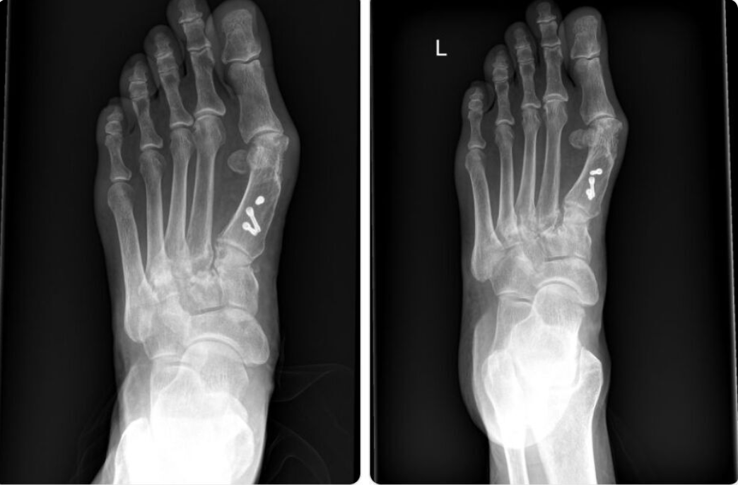
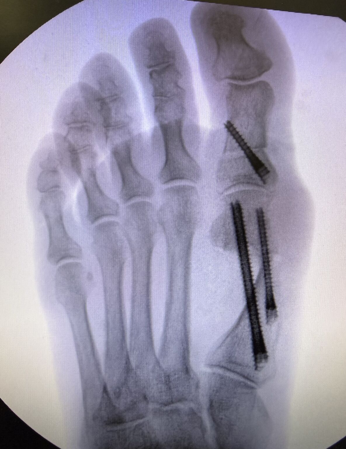
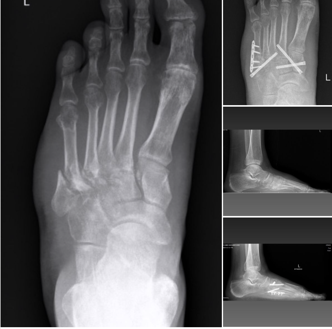
These are the X rays of a 68 year-old active female who underwent SCARF osteotomy for hallux valgus at another institution. How would you treat❓ About 1 year after surgery, she developed progressive foot pain without any history of new trauma. She underwent multiple consultations and several nerve block injections, but her symptoms continued. ▪️Now, 4 years after the initial surgery, she presents with severe, debilitating foot pain and marked restriction of motion, around the 1st metatarsophalangeal joint. ?Her pain progressed significantly, especially around the midfoot, and for the last 6 months she has required elbow crutches for walking. ❓ How would you approach this case? Thomas A. McDonald, MD FAAOS Tyler Gonzalez, MD, MBA, FAAOS Luis Sánchez-Robles, DPM, FACFAS, FAMOS, FASPS Walter Strash DPM, FACFAS Alan Yan Paulo Carvalho MD, PhD Pavel Mukherjee Dr. Marque A. Allen,DPM, FACFAS Jan S. Sean Campbell, MD Jordan Sisto, DPM, FACFAS Richard Freeman #RevisionSurgery #FootAndAnkleSurgery #OrthopaedicSurgery See original post on LinkedIn
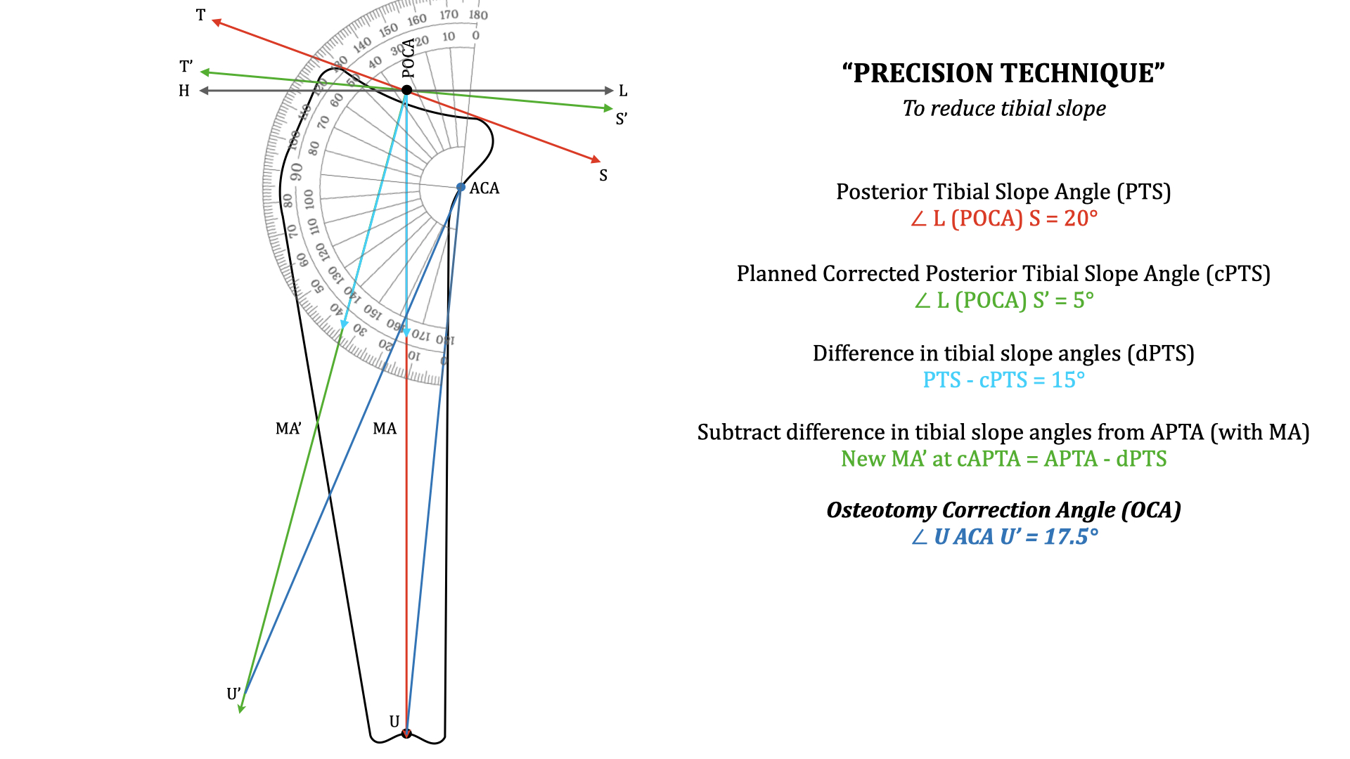
A novel “Precision technique” for preoperative planning of tibial slope correction osteotomy. High Tibial Osteotomy and Tibial Slope Correction Osteotomy requires precise preoperative planning. Tibial slope measurement and a slope correction osteotomy therefore requires planning without errors. Currently utilised preoperative planning techniques for slope correction are not very accurate. These can lead to errors in surgical correction and therefore impact the results. This novel “Precision technique” of planning overcomes errors in planning of the current traditional technique. It gives us the precise osteotomy correction angle by a simple measuring technique. The open access article which is attached highlights and gives all details of this novel technique. It can help one plan precisely, for accurate corrections to get the desired results. Link to the article - https://lnkd.in/gPuwrymD
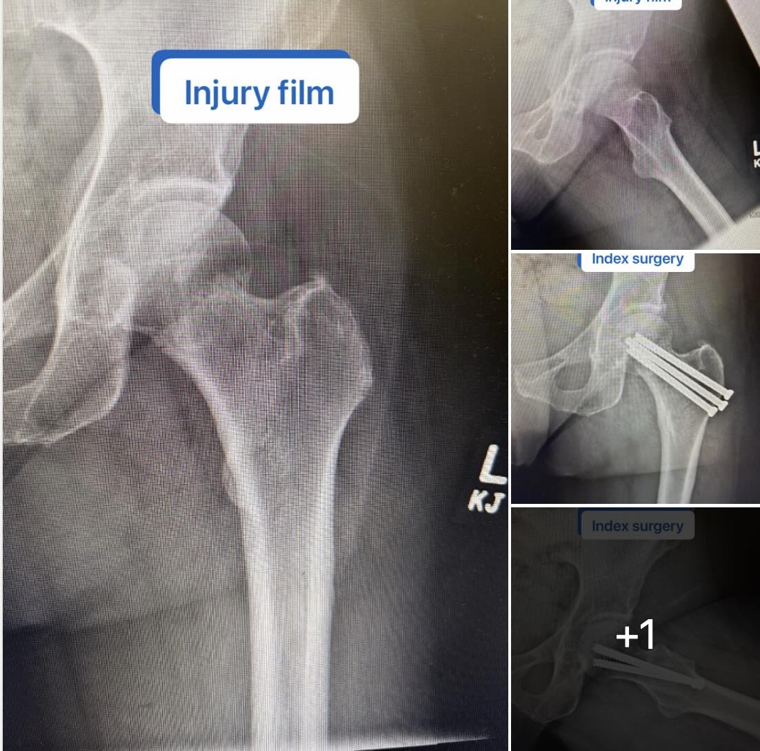
Lateral Hip Pain After Femoral Neck Fixation: What’s the Next Step? A patient presented after successful healing of an impacted femoral neck fracture treated by outside surgeon 5 years ago with closed reduction and percutaneous pinning. The fracture is healed—but now they’re dealing with persistent lateral hip pain. On exam: focal tenderness over the lateral screw heads. This is a scenario many of us see. Read more
❓For Lisfranc injury in younger patients, which fixation method do you prefer, transarticular screws with early removal, or a bridging plate construct?
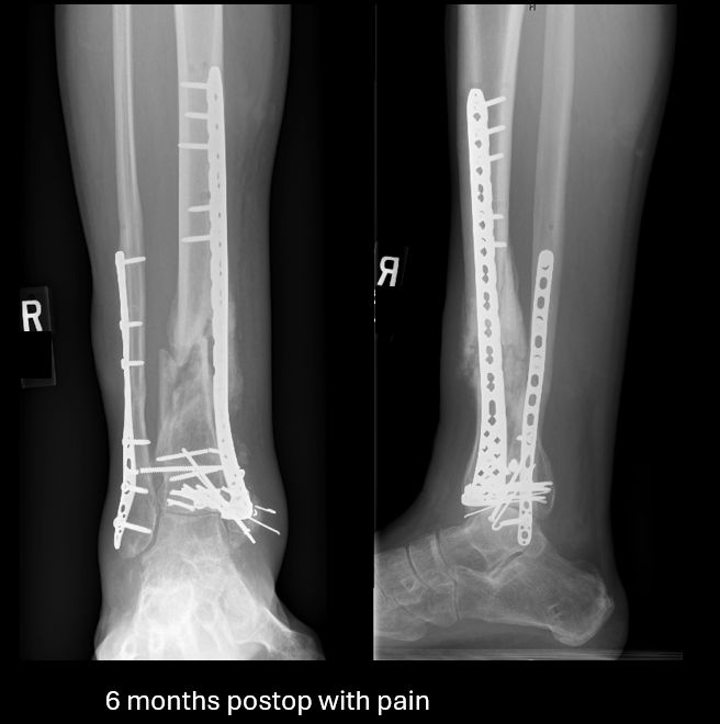
This young patient was 6 months out from my treatment of an open pilon fracture, presenting with pain and swelling. The soft tissues were in good condition. There were no signs of infection and labs were normal. Images showed callus but also loss of coronal alignment, nonunion, and broken implants. The injury and immediate postop films are available for review. Step 1: Is this an extra-articular or intra-articular problem? This one has a healed joint and is an extra-articular issue. Step 2: Mechanical or biologic problem? Primarily a mechanical problem. - Uni-columnar bridge plate likely insufficient to last for the entire duration of healing. - Progressive loss of coronal alignment worsened the environment The biology was affected by initial injury but presence of callus and hypertrophic nonunion suggests it is not the dominant issue Treatment strategy: - Remove failed implants - Restore alignment with a medially based frame Stabilize with medullary nail Reaming stimulated the biology, but the key intervention is mechanical optimization This led to reliable healing in this case. In my experience, medullary nailing with realignment is an excellent option for hypertrophic extra-articular pilon nonunion. What are your thoughts on this problem? Do you like medullary nailing? Any other good treatment options?
A case that highlights one of the most important principles in knee arthroplasty: patient selection matters. I recently treated a 60-year-old male who presented one year after a medial unicompartmental (partial) knee replacement performed elsewhere. He continued to have significant pain and functional limitation. On evaluation, he had “progressive” arthritis in the lateral and patellofemoral compartments—ultimately requiring conversion to a total knee arthroplasty. I suspect he had significant changes prior to his partial knee arthroplasty, but he was unable to obtain preoperative images. Partial knee replacement can be an excellent operation: Faster recover, More normal knee kinematics, Preservation of bone and ligaments But it is not forgiving when indications are stretched. The key principle: Partial knee replacement is best reserved for patients with truly isolated unicompartmental disease. When arthritis is already present—or likely to progress—in other compartments, the risk of early failure and revision increases significantly. In this case, the patient did well after conversion to a total knee, but it underscores an important point: The success of a partial knee begins before the incision—with proper patient selection. As surgeons, the decision is not just what we can do, but what we should do to give patients the best long-term outcome. Click here to view the original post
Would you jump from a plane with only one parachute & no reserve parachute?! Not me. Similarly, I’m traditionally a 2-screw fixation surgeon for my first metatarsal during MIS bunion surgery. Maybe 1 screw could get the job done in certain situations & you could get lucky, but why take the risk & have to deal with potential complications if that 1 screw fails? Steps made simple: 1. Place reference wire at osteotomy site. 2. Targeter places wires in perfect position. 3. Make transverse osteotomy. 4. Employ “crow-bar” for 3D deformity correction. 5. Advance wires & place fixation. Do you use a reserve parachute? Click here to view the original post
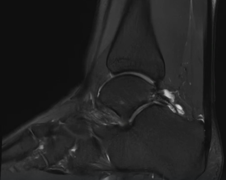
Posterior ankle pain in athletes isn’t always “just impingement.” Often it’s a combined os trigonum + FHL pathology problem. Takeaway from Arthrex TPC Meeting Recent patient with persistent posterior ankle pain—worse with push-off and plantarflexion. Workup: ▪️ Os trigonum on imaging ▪️ Pain with resisted hallux flexion → FHL involvement ▪️ Failed nonoperative management Surgical Decision: Arthroscopic vs Open Arthroscopic approach ✔ Less soft tissue disruption ✔ Faster recovery ✔ Direct visualization of posterior ankle Open approach ✔ Useful for large ossicles or abnormal anatomy ✔ Better for extensive scarring or tenosynovitis ✔ More direct control of the FHL tendon Critical Point If you remove the os trigonum and ignore the FHL… you will miss the primary pain generator in some patients. In this case: ▪️ Posterior ankle arthroscopy ▪️ Os trigonum excision ▪️ FHL tenolysis + release Let’s Talk Technique (for those doing open) Curious how others are approaching this: Medial vs lateral approach? Medial: more direct access to FHL, but tighter working space Lateral: more familiar for os trigonum, but indirect access to FHL Takeaway Posterior ankle impingement is often a two-structure problem Bone + tendon If you’re not evaluating both… you’re not done. Click here to view the original post
How are you managing failed total ankle replacement? This patient underwent an ankle replacement 5 years ago at an outside hospital. Patient has been in pain and walking on the side of their foot. Slowly getting worse. Clinical Varus. Radiohraphic varus. Infinity tibia and Inbone talus What’s next? - Conversion to fusion - Revision to a replacement - Staged revision Patient underwent removal of implants then revision to Inbone tibia and Invision talus. Frozen & GS negative. Foot deformity needed to be corrected as well. Dwyer and lateral displacement calcaneal osteotomy with base of 1st DF Osteotomy Lateral ligaments stretched out. Modified Brostrum was needed too What would you have done differently? Click here to view the original post
You're seeing a preview. Sign in to see all posts and join the Surgzon community.
Sign in to see more