
Biomechanical Perspective of Cavovarus Reconstruction
Cavovarus reconstruction is often taught as a series of procedures — calcaneal osteotomy, tendon transfers, first metatarsal dorsiflexion osteotomy, and occasionally midfoot procedures.
But it may be more useful to think about the problem from a biomechanical perspective first.
The goal of surgery is to restore hindfoot alignment and reduce the talonavicular joint, which re-establishes the normal relationship between the rearfoot and midfoot. Once that occurs, the true residual forefoot deformity becomes apparent.
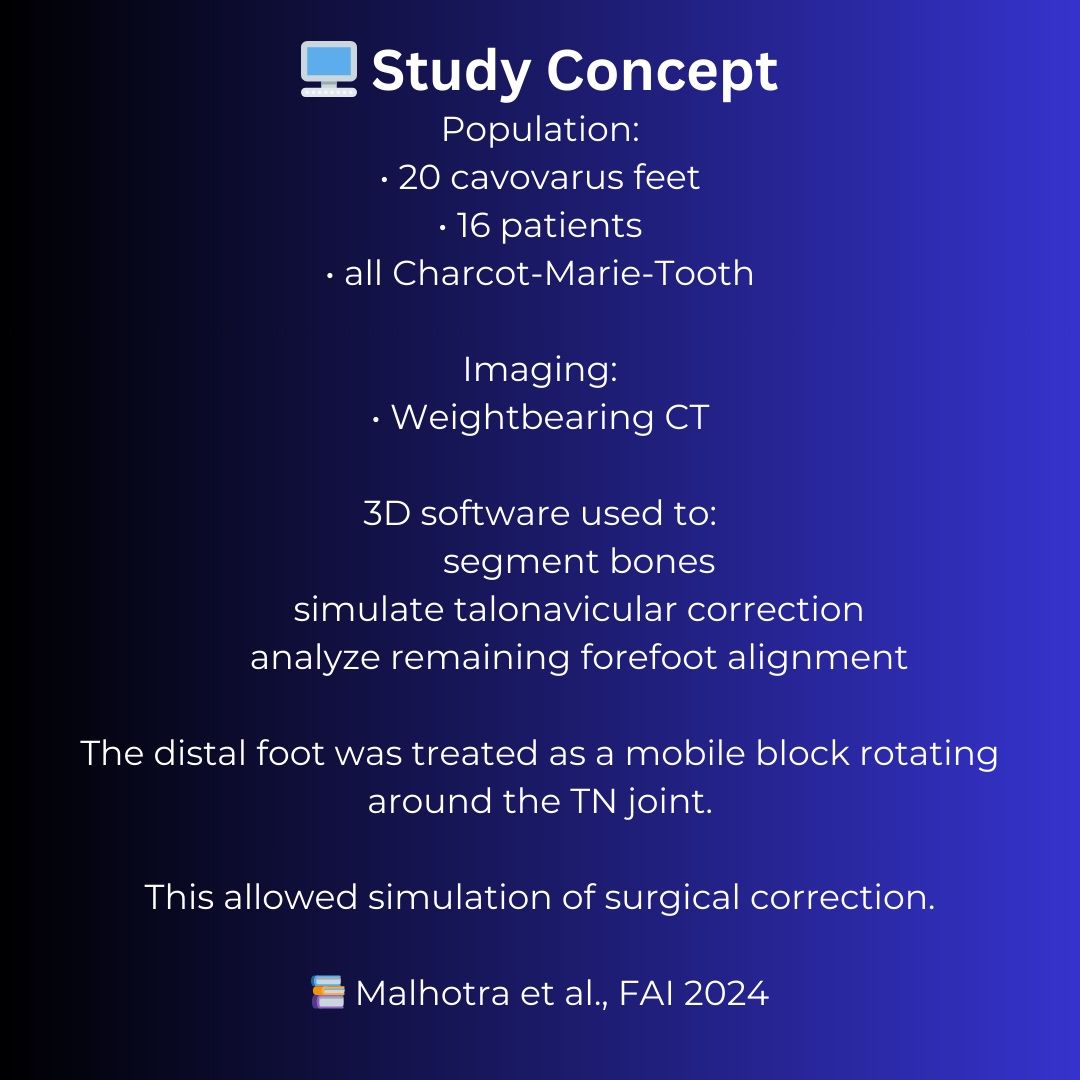
This study used weightbearing CT modeling to simulate talonavicular correction in cavovarus feet and identified four predictable forefoot morphotypes that remain after hindfoot correction.
The most common pattern was isolated plantarflexion of the first ray, which explains why a 1st metatarsal dorsiflexion osteotomy is such a common component of cavovarus reconstruction.
From a biomechanical perspective, this makes sense. According to Kirby’s rotational equilibrium theory, the foot assumes a position during weightbearing where pronation and supination moments around the subtalar joint axis are balanced. Changes in the relationship between the subtalar axis and the forefoot alter these moment arms and influence how ground reaction forces are distributed across the foot.
Once hindfoot alignment is restored, the remaining forefoot deformity represents the mechanism through which the foot is attempting to maintain that equilibrium.
Understanding these patterns may help surgeons move beyond generic cavovarus algorithms and toward more precise forefoot balancing strategies that restore a stable tripod and a durable plantigrade foot.